Altered ocular microvasculature in patients with systemic sclerosis and very early disease of systemic sclerosis using optical coherence tomography angiography
Por um escritor misterioso
Descrição

Prevalence of ocular findings.

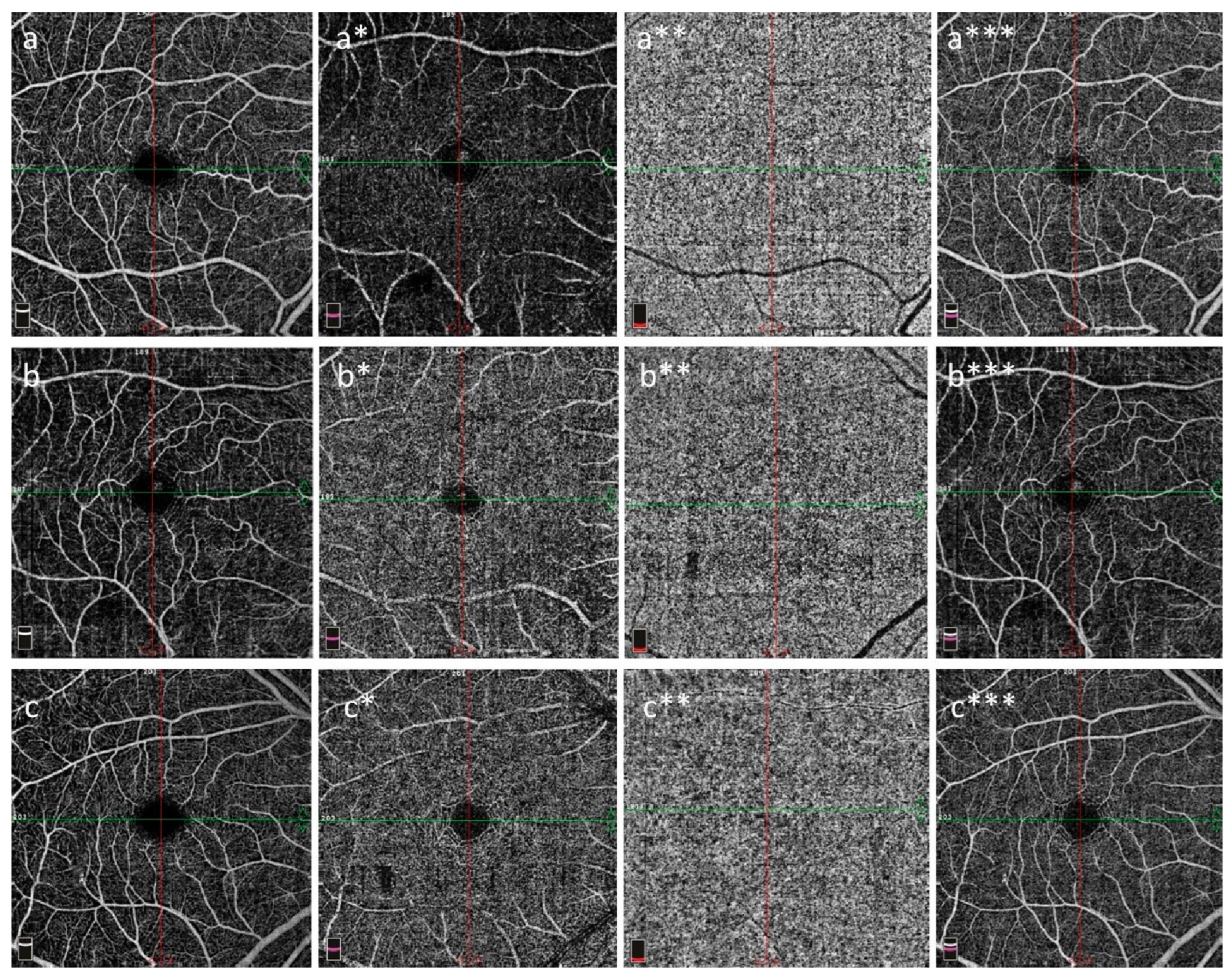
A1–C2) Three exemplary 3 × 3 mm² en-face optical coherence tomography

Optical coherence tomography angiography findings in Williams-Beuren syndrome

Optical coherence tomography angiography - ScienceDirect

Early alterations in retinal microvasculature on swept-source optical coherence tomography angiography in acute central serous chorioretinopathy

Optical coherence tomography: From technology to applications in ophthalmology - Everett - 2021 - Translational Biophotonics - Wiley Online Library
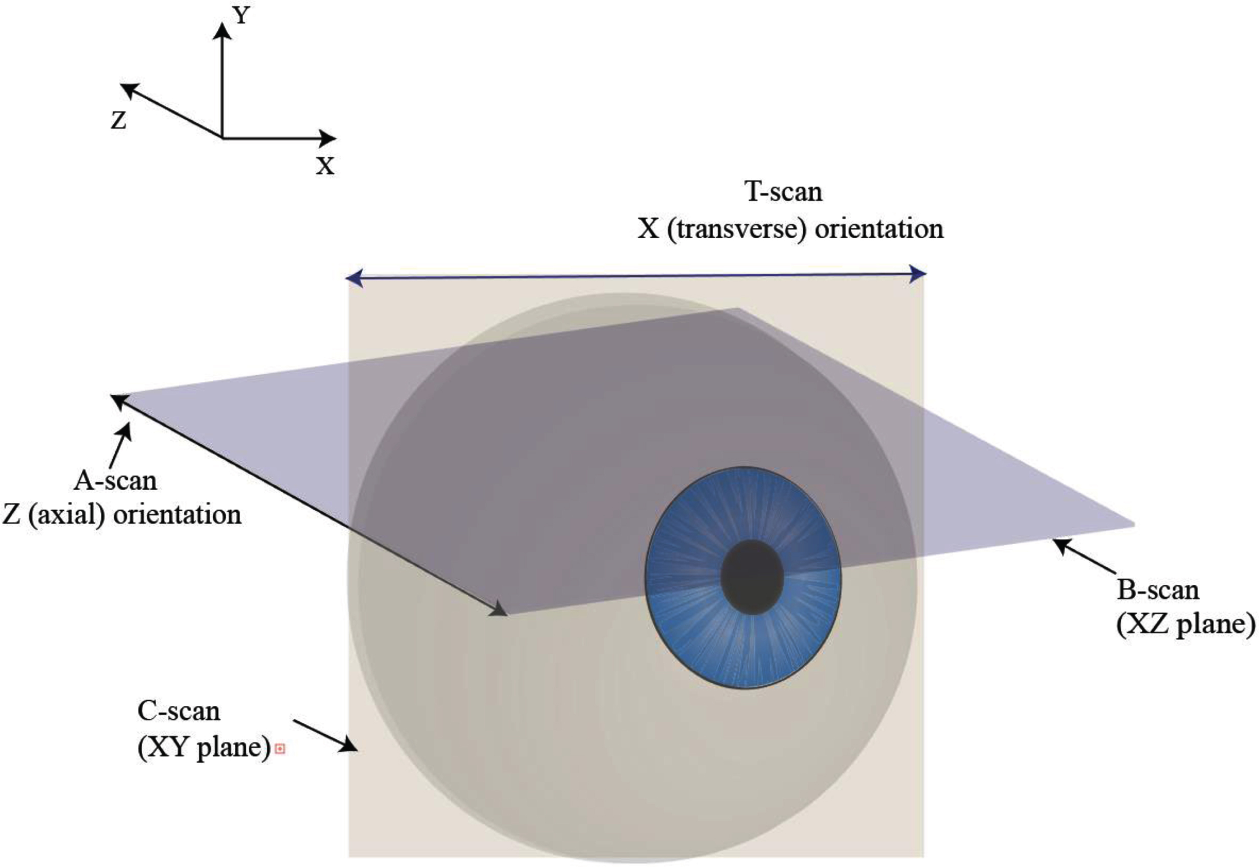
Optical Coherence Tomography Angiography (OCTA) of the eye: A review on basic principles, advantages, disadvantages and device specifications - IOS Press

Representative optical coherence tomography angiography (OCTA) images
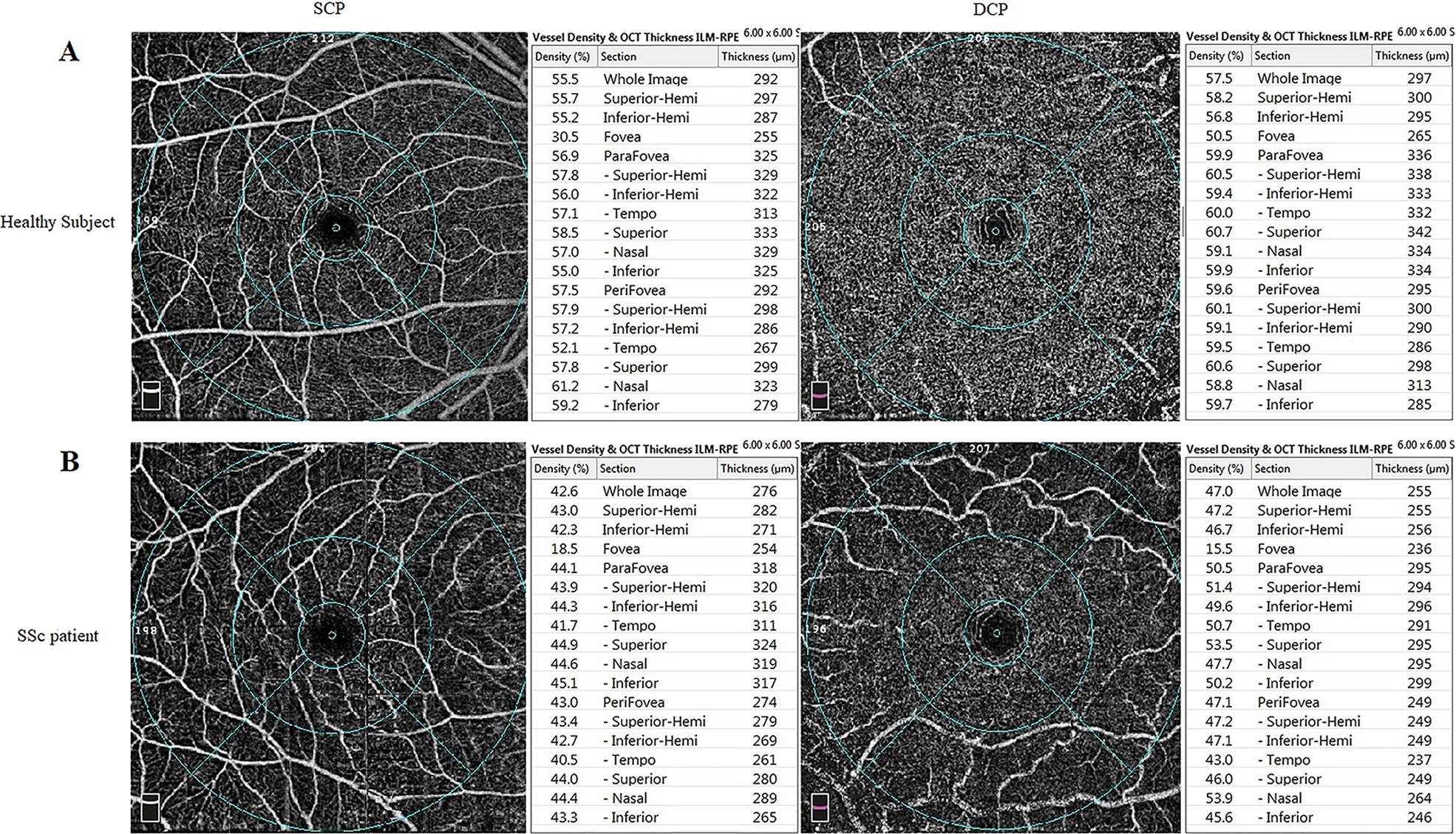
Analysis of retinal and choroidal microvasculature in systemic sclerosis: an optical coherence tomography angiography study

JPM, Free Full-Text

The Function of Retinal Thickness and Microvascular Alterations in the Diagnosis of Systemic Sclerosis

Optical coherence tomography angiography - ScienceDirect
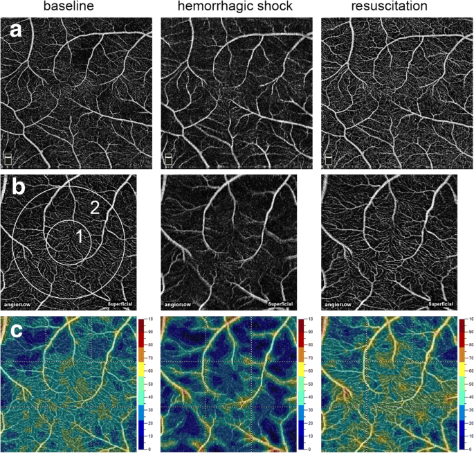
Feasibility of optical coherence tomography angiography to assess changes in retinal microcirculation in ovine haemorrhagic shock, Critical Care
Diagnostics, Free Full-Text

Optical coherence tomography angiography - ScienceDirect


