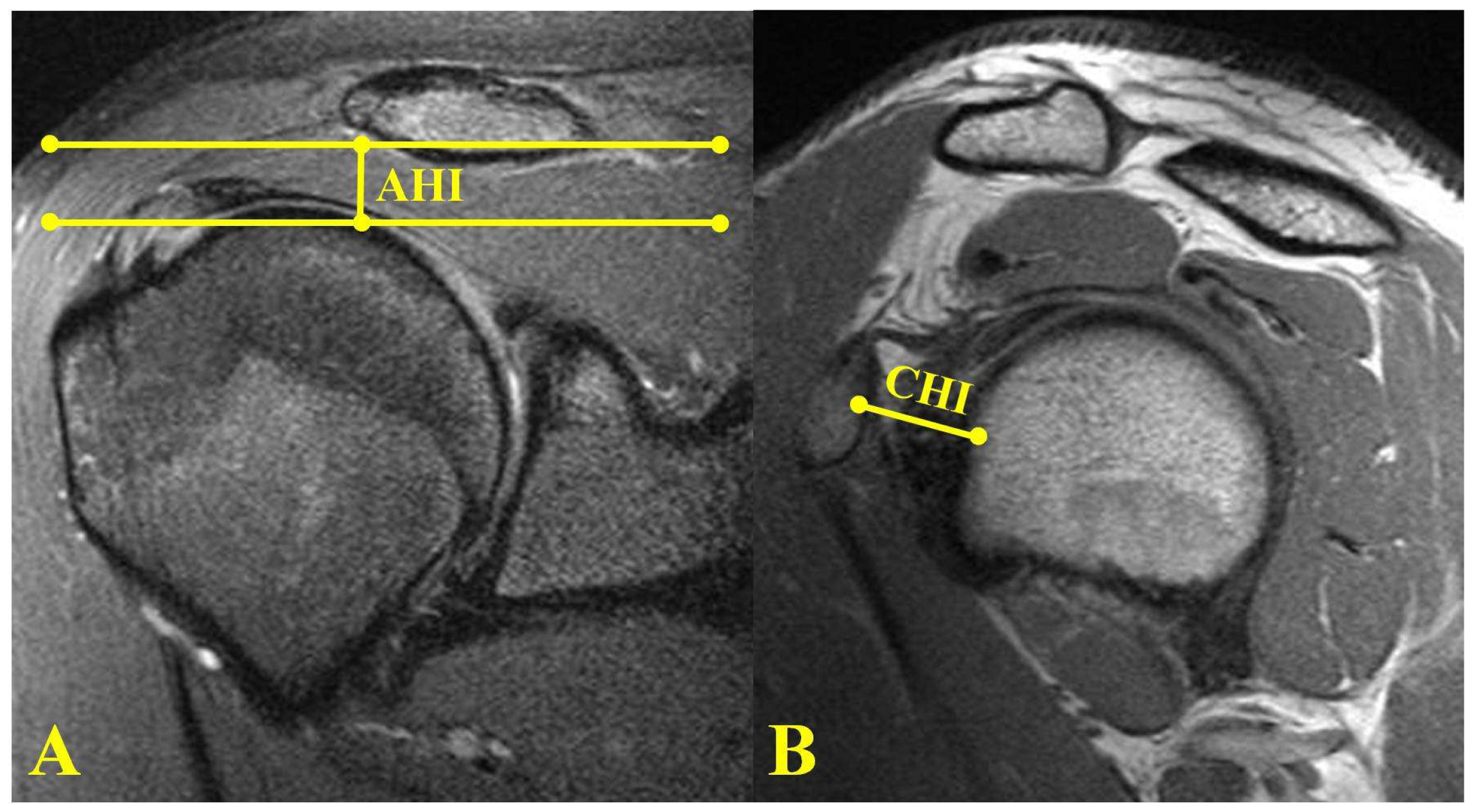
Typical magnetic resonance imaging scan showing the coracohumeral
Por um escritor misterioso
Descrição

Typical magnetic resonance imaging scan showing the coracohumeral

JCM, Free Full-Text

Diagnostics, Free Full-Text

Presentation1, radiological imaging of adhesive capsulitis(frozen shoulder).

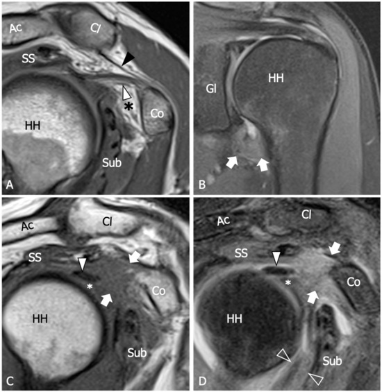
Spectrum of lesions of the acromioclavicular joint: imaging features

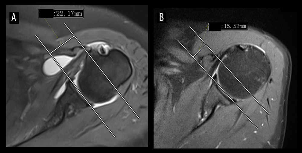
Medical Science Monitor Identification of Diagnostic Magnetic Resonance Imaging Findings in 47 Shoulders with Subcoracoid Impingement Syndrome by Comparison with 100 Normal Shoulders - Article abstract #936703

The Rotator Interval: A Review of Anatomy, Function, and Normal and Abnormal MRI Appearance

Spectrum of Various Morphological Changes Detected on High Resolution Ultrasound & MRI in Patients with Painful Shoulder

Pain related to rotator cuff abnormalities: MRI findings without clinical significance - Bencardino - 2010 - Journal of Magnetic Resonance Imaging - Wiley Online Library


