Advanced Quantitative Fluorescence Microscopy to Probe the Molecular Dynamics of Viral Entry, Science Lab
Por um escritor misterioso
Descrição
Viral entry into the host cell requires the coordination of many cellular and viral proteins in a precise order. Modern microscopy techniques are now allowing researchers to investigate these interactions with higher spatiotemporal resolution than ever before. Here we present two examples from the field of HIV research that make use of an innovative quantitative imaging approach as well as cutting edge fluorescence lifetime-based confocal microscopy methods to gain novel insights into how HIV fuses to cell membranes and enters the cell.

NgR1 binding to reovirus reveals an unusual bivalent interaction and a new viral attachment protein

Beyond mass spectrometry, the next step in proteomics

Peptide-Induced Fusion of Dynamic Membrane Nanodomains: Implications in a Viral Entry

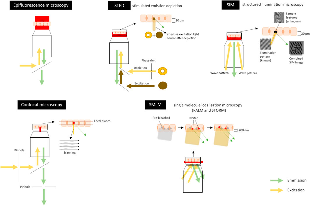
Frontiers Microscopic Visualization of Cell-Cell Adhesion Complexes at Micro and Nanoscale

Phasor S-FLIM workflow and robustness to noise of the phasor

2021 Top 10 Innovations The Scientist Magazine®

Altered host protease determinants for SARS-CoV-2 Omicron

A chimeric virus-based probe unambiguously detects live circulating tumor cells with high specificity and sensitivity - ScienceDirect

Advancements in detection of SARS-CoV-2 infection for confronting COVID-19 pandemics - Laboratory Investigation


