A high magnification image of synapse obtained by electron microscopy
Por um escritor misterioso
Descrição

Choosing the Right Scanning Electron Microscope for Your

Modern field emission scanning electron microscopy provides new

Figure 1, Electron microscope images of worm synapses - WormBook

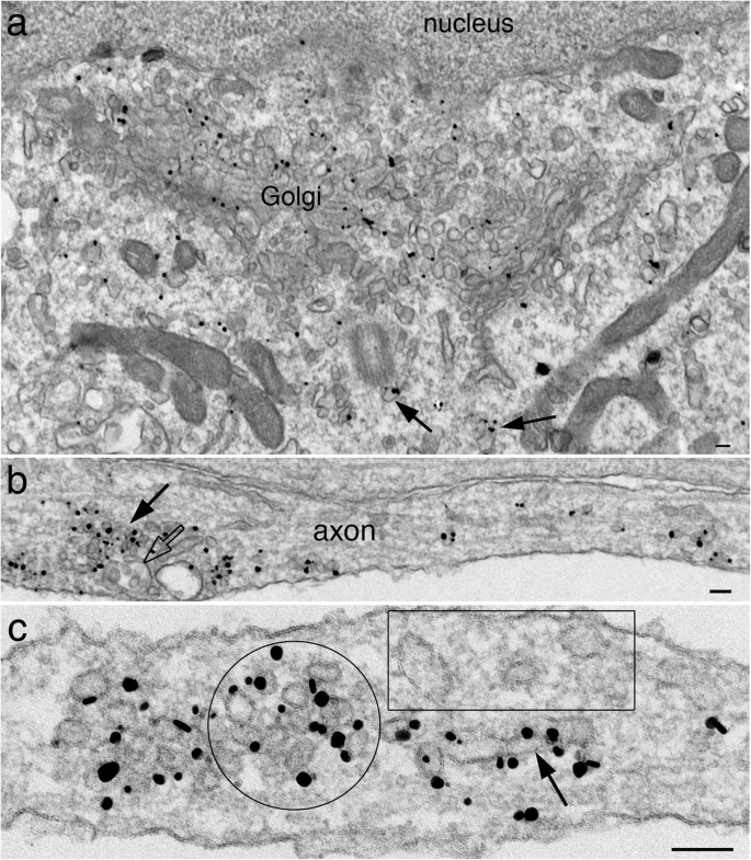
Immunogold labeling of synaptic vesicle proteins in developing

FSFF Human Nerve Synapse Model, The Magnified Structure of The

Figure 2 from History of the Electron Microscope in Cell Biology

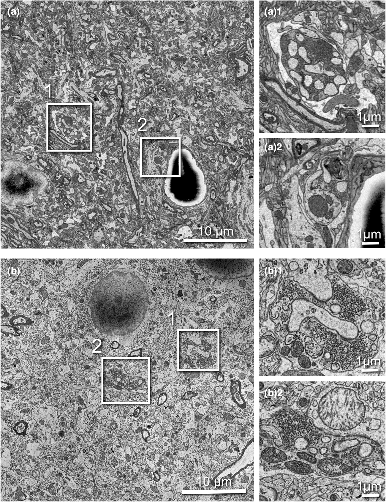
Differentiation and Characterization of Excitatory and Inhibitory

Targeting Functionally Characterized Synaptic Architecture Using

High-resolution volumetric imaging constrains compartmental models

Functional Electron Microscopy, “Flash and Freeze,” of Identified

Role of Electron Microscopy in Tumor Diagnosis: A Review

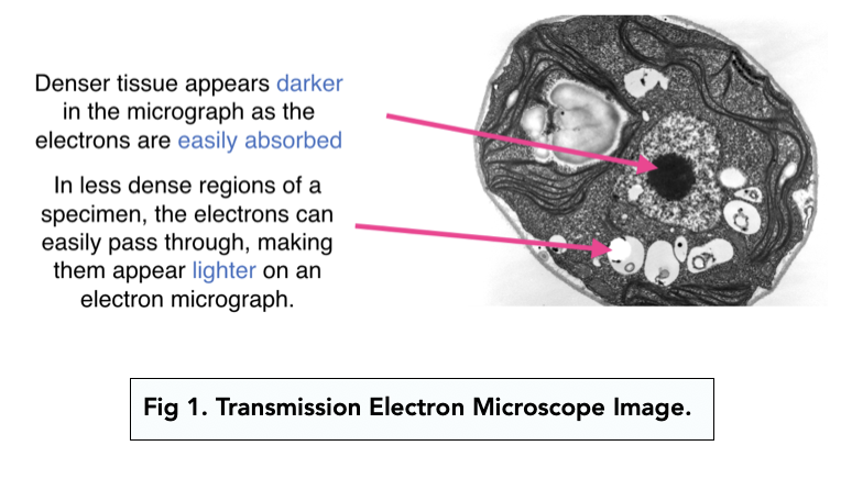
Studying Cells: Electron Microscopes (A-level Biology) - Study Mind

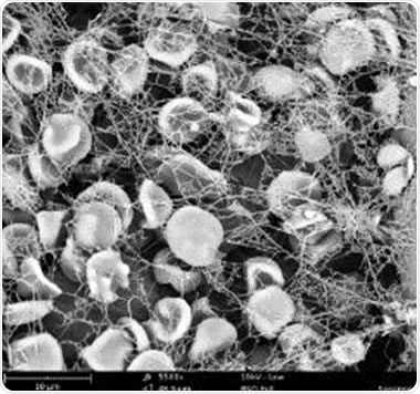
Phenom Scanning Electron Microscope Accelerates Blood Clotting


